Does Alcohol Kill Brain Cells? Part Two
This week, Kenneth Anderson, MA, explores MRI and brain shrinkage research in part 2 of the series, “Does Alcohol Kill Braincells?” Part 1 can be found here.
By Kenneth Anderson, MA
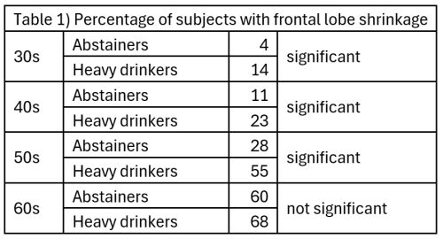
 A 2001 study by Kubota et al. performed MRIs on 1,432 employees and family members (1,061 men and 371 women) of a large Japanese corporation to investigate shrinkage of the frontal lobe of the brain. Subjects were each categorized by their drinking status (abstainers, light drinkers, moderate drinkers, heavy drinkers), age (30s, 40s, 50s, or 60s), and brain shrinkage (shrunken, not shrunken). Heavy drinkers were classified as those who drank 350 grams of ethanol (25 US standard drinks) or more per week. There were no significant differences between the abstainers, light drinkers, and moderate drinkers in terms of brain shrinkage; however, significantly more heavy drinkers showed brain shrinkage compared to abstainers. Table 1 shows the percentage of subjects in each age group with frontal lobe shrinkage for abstainers and heavy drinkers. For subjects in their 30s, 4% of abstainers showed frontal lobe shrinkage and 14% of heavy drinkers showed frontal lobe shrinkage, etc.
The researchers concluded that, although many factors, including age and heavy drinking, contributed to shrinkage of the frontal lobe, the greatest factor was age (accounting for 30.0% of variance), followed by heavy drinking (accounting for 11.3% of variance). The researchers also concluded that the reason there was no significant difference between abstainers and heavy drinkers in their 60s was that many older subjects had moderated their drinking. Since this is a cross-sectional study, we can’t use it to infer causality. In other words, we can’t tell if drinking caused frontal lobe shrinkage or if frontal lobe shrinkage caused drinking.
Ding et al. (2004) conducted an MRI study of 1909 middle-aged adults. Subjects were divided into five categories based on their drinking habits: never drinkers, ex-drinkers, occasional drinkers (less than one US standard drink per week), low drinkers (one to six US standard drinks per week, and high drinkers (seven or more US standard drinks per week). The MRI was used to measure the size of the grooves (sulci) on the surface of the brain and the size of the ventricles (hollow cavities which contain cerebrospinal fluid) found within the brain. Larger grooves indicate brain shrinkage, as do larger ventricles. The size of the grooves was graded 0 to 9; a zero representing the normal size and a 9 representing the largest size. The same grading system was used for the ventricles. It was found that groove (sulci) size increased by 0.009 grade for each additional drink consumed per week. Likewise, ventricle size increased by 0.01 grade for each additional drink consumed per week. The differences for both groove (sulci) size and ventricle size were significant, indicating brain shrinkage proportional to the number of drinks consumed per week, even in moderate drinkers.
A 2001 MRI study of 3,660 adults aged 65 years and older by Mukamal et al. found similar results. Compared to lifelong abstainers, heavy drinkers (more than 14 US standard drinks per week) showed significant increases in the size of the brain’s grooves and ventricles, indicating brain shrinkage. Changes in grooves and ventricles were graded on a scale of 0 to 9; the difference between lifelong abstainers and heavy drinkers was 0.2 grades.
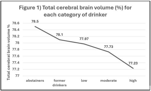
 Paul et al. (2008) conducted an MRI study of 1,839 subjects (861 males and 978 females) aged 33 to 88. Subjects were classified as lifelong abstainers, former drinkers, low drinkers (one to seven US standard drinks per week), moderate drinkers (eight to 14 US standard drinks per week), and high drinkers (15 or more US standard drinks per week). Paul et al. compared the total cerebral brain volume (TCBV) of each of these groups to see what effect alcohol had on total cerebral brain volume. Total cerebral brain volume was calculated by finding what percentage of the skull was occupied by the brain, and hence, was expressed as a percentage. Total cerebral brain volume was adjusted for age, sex, body mass index (BMI) and Framingham Stroke Risk Profile (FSRP). Figure 1 shows the results of this comparison. High drinkers showed a significantly smaller total cerebral brain volume than all four other groups (high drinkers vs. abstainers p < 0.01; vs. former p < 0.01; vs. low p < 0.01; vs. moderate p = 0.045). Moderate drinkers had a significantly smaller total cerebral brain volume than former drinkers. High drinkers had about 1.3% less total cerebral brain volume than lifelong abstainers. Females showed a significantly greater effect of alcohol on total cerebral brain volume than males.
However, a 2014 MRI study of 589 subjects aged 65 and over by Gu et al. found a significantly larger total cerebral brain volume for light and moderate drinkers compared to past-year abstainers (i.e., those reporting that they had drunk no alcohol during the past year).
A 2004 MRI study by den Heijer et al. found that light to moderate drinking was protective against damage to white matter in the brain. This study also found that light to moderate drinking was protective against shrinkage of the hippocampus and amygdala in subjects with the apolipoprotein (APOE) epsilon 4 allele, but not in other subjects. There were 1,074 subjects in this study, aged 60 to 90. None had dementia.
Sachdev et al. (2008) conducted an MRI study of 383 Australian subjects (211 men and 172 women) aged 60 to 64 using voxel-based morphometry. “Voxel” is a portmanteau word made by combining the words “volume” and “pixel.” A voxel is essentially a three-dimensional pixel used to measure volume. Voxel-based morphometry uses a computer to make a three-dimensional model of the brain based on the MRI scan information. Voxel-based morphometry allows for a much more precise comparison of the volumes of various areas of the living brain than earlier methods did. Subjects were low to moderate drinkers: men drank an average of 6.6 US standard drinks (9.30 Australian units) per week and women drank an average of 3.02 US standard drinks (4.26 Australian units) per week. Only 13 men averaged more than 2.84 US standard drinks (4 Australian units) per day, and only seven women averaged more than 1.42 US standard drinks (2 Australian units) per day. Men were found to have more gray matter and less white matter in certain brain areas the more they drank: this relationship was linear. No effect of alcohol consumption on gray or white matter was found for women.
Taki et al. (2004) carried out an MRI study of 769 Japanese subjects aged 16 to 79 (356 men and 413 women) using voxel-based morphometry. Subjects were classified as low drinkers (0 to one drinking day per week) or high drinkers (more than one drinking day per week). Among male subjects, high drinkers showed significantly less gray matter but significantly more white matter than low drinkers. Among female subjects, there were no significant differences due to drinking status; however, female subjects consumed little alcohol compared to male subjects.
De Bruin et al. (2005) conducted an MRI study of 91 Dutch subjects (47 male and 44 female) using voxel-based morphometry. Abstainers were excluded from this study, as were people with alcohol dependence. Current and lifetime alcohol consumption was measured. Current alcohol consumption had no effect on gray or white matter; however, lifetime alcohol consumption affected both gray and white matter. Male subjects had an average lifetime alcohol intake of 240 kilograms of alcohol. Female subjects had an average lifetime alcohol intake of 170 kilograms. Lifetime alcohol consumption had no effect on gray or white matter for female subjects. However, male high lifetime drinkers showed significantly less gray matter and significantly more white matter in certain brain areas than male low lifetime drinkers. For example, a male high lifetime drinker who drank an average of seven Dutch alcohol units per week over his lifetime had a gray-matter density in his right frontal gyrus which was 28% lower than the male consuming an average of one Dutch alcohol unit per week over his lifetime. Likewise, a man consuming seven Dutch alcohol units per week over his lifetime had an 11% higher white-matter density in his right frontal gyrus, a 19% lower gray-matter density and a 34% higher white matter in his right parietal region as compared to a man drinking one Dutch alcohol unit per week over his lifetime. These are the brain areas associated with visual attention. (Note: this article is unclear as to whether one Dutch alcohol unit is 10 gram of ethanol or 12 grams; the US standard drink is 14 grams.)
Sasaki et al. (2009) conducted an MRI study of 211 Japanese subjects (114 males and 97 females) on the impact of lifetime alcohol consumption on the brain using voxel-based morphometry. Subjects ranged in age from 21 to 72 years old with an average age of 37. Lifetime alcohol consumption for males ranged from 0 to 1,551 kilograms of ethanol with an average of 143 kilograms. For females, lifetime consumption ranged from 0 to 397 kilograms of ethanol with an average of 32 kilograms. For males, the researchers found no impact of lifetime alcohol consumption on the total volume of either gray matter or white matter. Among females, the only impact of alcohol found by the researchers was an increase in the mean diffusivity of the right amygdala. The amygdala is associated with fear, emotions, and motivation.
Fukuda et al. (2009) conducted an MRI study of 385 rural Japanese subjects (149 men and 236 women) over the age of 40; the average age was 67. Subjects were categorized as non-drinkers, light drinkers, and moderate drinkers. The category non-drinker included lifelong abstainers, former drinkers, and anyone who drank less than once a month. The category light drinker included those who drank at least once a month up to those who drank less than 70 grams of ethanol (five US standard drinks) per week. The category moderate drinker included everyone who drank 70 or more grams of ethanol (five US standard drinks) per week. About half of the moderate drinkers (58) consumed 140 grams of ethanol (10 US standard drinks) or more per week. The researchers measured brain shrinkage by calculating the percentage of the area of the skull occupied by an MRI slice of the brain. (Note that this differs from the 2008 study by Paul et al. which calculated the percentage of volume of the skull occupied by the brain. This study looked at area instead of volume.) The percentage of skull area occupied by the brain for each drinker category was as follows: non-drinkers, 92.1%; light drinkers, 91.4%; and moderate drinkers, 90.8%. The difference between non-drinkers and moderate drinkers was statistically significant, indicating brain shrinkage among moderate drinkers.
Anstey et al. (2006) conducted an MRI study of 385 Australian adults (211 men and 174 women) aged 60 to 64. Subjects were chosen at random from the Australian electoral rolls. Men who drank more than 280 grams of alcohol (20 US standard drinks) per week, and women who drank more than 140 grams (10 US standard drinks), were classified as heavy drinkers. Everyone else was classified as a light drinker. For men, heavy drinkers showed significantly more gray matter volume than light drinkers, but significantly less white matter volume. For women, there was no difference in gray matter volume, but heavy drinking women showed significantly less white matter volume. There were no alcohol-related differences found in the hippocampus (the seat of memory) or the corpus callosum (the bridge between the right and the left halves of the brain).
Topiwala et al. (2017) conducted an MRI study of 527 UK adults (424 men and 103 women) using voxel-based morphometry. Although alcohol consumption and other variables had been tracked for these subjects for 30 years, the MRIs were conducted only once, at the end of the study, making this a cross-sectional study. Subjects were divided into six groups based on alcohol consumption: abstainers (0 to 8 grams of alcohol, i.e., 0 to 0.6 US standard drinks, per week), light drinkers (8 to 56 grams, i.e., 0.6 to 4 US standard drinks, per week), light-moderate drinkers (56 to 112 grams, i.e., 4 to 8 US standard drinks, per week), moderate drinkers (112 grams to 168 grams i.e., 8 to 12 US standard drinks, per week), moderate-heavy drinkers (168 grams to 240 grams, i.e., 12 to 17 US standard drinks, per week), and heavy drinkers (168 grams or more, i.e., 17 or more US standard drinks, per week). It was found that the odds ratio of having shrinkage of the hippocampus increased with the amount of alcohol consumed in a dose-dependent manner. For the right hippocampus, the odds ratios of shrinkage were as follows: abstainers, 1 (this is the baseline, i.e., a value of “1” means no increase in the odds); light drinkers, 1.5; light-moderate drinkers, 2.0; moderate drinkers, 3.4; moderate-heavy drinkers, 3.6; and heavy drinkers, 5.8. In other words, the heavy drinkers were 5.8 times more likely than abstainers to have shrinkage of the right hippocampus. For the left hippocampus, the odds ratios of shrinkage were as follows: light drinkers, 1.3; light-moderate drinkers, 1.4; moderate drinkers, 1.9; moderate-heavy drinkers, 1.9; and heavy drinkers, 5.7. Topiwala et al. also found that increased alcohol consumption led to reduced gray matter density and reduced white matter microstructural integrity.
Fein et al. (2002) conducted an MRI study of 24 men with alcohol dependence who had never been to alcohol treatment and who were not seeking treatment; this sample was compared to 17 men who were light drinkers who served as controls. The alcohol dependent group met DSM-IV criteria for alcohol dependence and consumed an average of 559 grams of alcohol (40 US standard drinks) per week, with an average lifetime consumption of 644 kilograms of alcohol (46,030 US standard drinks). The light drinking group consumed an average of 72 grams of alcohol (5 US standard drinks) per week, with an average lifetime consumption of 46.4 kilograms of alcohol (3,315 US standard drinks). Initial MRI data were confounded by the fact that the sample was not age matched; therefore, the older alcohol dependent subjects were removed to create an age matched sample consisting of 16 men with alcohol dependence and 17 men who were light drinkers. In the age-matched sample, the only significant difference between the alcohol dependent group and the control group was a reduction in gray matter in the alcohol dependent group in the posterior prefrontal cortex (which is involved in basic executive processes) and the dorsolateral prefrontal cortex (which is involved in working memory, cognitive flexibility, planning, inhibition, and abstract reasoning). There were no significant differences in total white matter or total gray matter between the two groups.
Daviet et al (2022) conducted a study which utilized MRI data from 36,678 middle-aged and older adults in the UK Biobank. The large number of subjects made this study very sensitive to significant effects. It was found that alcohol consumption accounted for only 1% of the variance total gray matter volume and 0.3% of the variance total white matter volume; however, both these figures were significant. Table 2 shows the reductions in total gray and total white matter in standard deviations (SDs) for each additional UK unit of alcohol (8 grams) consumed per day. Going from 0 UK units (0 US standard drinks) per day to 4 UK units (2.3 US standard drinks) per day leads to a reduction in total gray matter of 0.577 standard deviations and a reduction in total white matter of 0.367 standard deviations.

All of these community-based studies were cross sectional, and hence, causality cannot be inferred. In part three, we will look at clinical samples.
Liked this article? Read part one here!
